Rib Cage Muscles Anatomy - Muscles of the thorax and abdomen. | Rib cage, Anatomy, Art - Collectively referred to as the rib cage costal cartilages sternum.
byAdmin-
0
Rib Cage Muscles Anatomy - Muscles of the thorax and abdomen. | Rib cage, Anatomy, Art - Collectively referred to as the rib cage costal cartilages sternum.. Contributing to their role in protecting the internal thoracic organs. It also functions as an attachment site for your respiratory muscles rib cage pain can arise from injury to any of the muscles, bones, nerves or joints within the thoracic cage region. Abdomen & ribs muscle movements. The thoracic cage refers to the skeleton of the thorax: Ribs are not merely armour for the organs inside our torsos, as we rib fractures are a common and very painful injury, with the middle ribs the most likely ones to get the muscles that move the ribcage itself are the intercostal muscles.
Muscular system anatomy:muscles of the thoracic cage torso model description. The thoracic cage refers to the skeleton of the thorax: In the anatomical position, the angles align with the medial border of the scapula. It provides a strong framework onto which the muscles of the cramps in ribcage are often observed in those who strain or overwork their upper body. They are more involved in forced expiration and coughing to forcibly shrink the chest and.
Anatomy-of-the-Human-Rib-Cage - Badass chiropractic and ... from drkarenhudes.ca Some of the most common causes. In the anatomical position, the angles align with the medial border of the scapula. • raise rib cage for inhaling & depresses rib cage for exhaling. Muscles that move the rib cage attach to the rib cage. See more ideas about anatomy, anatomy study, rib cage anatomy. Rib cage anatomy and breathing. Des milliers de nouvelles images de grande qualité ajoutées chaque jour. These muscles may be located anteriorly, posteriorly, and/or laterally.
Learn anatomy faster and remember everything you learn.
Ribs & thoracic cage muscles attachments. Contributing to their role in protecting the internal thoracic organs. Various skeletal muscles are attached to the rib cage. The rib cage, shaped in a mild cone shape and more flexible than most bone sets, is made up of varying elements such as the thoracic vertebra, 12 equally paired ribs, costal cartilage, and held together anteriorly by the sternum. Posterior rib cage muscles : The rib cage is a primarily protective structure, encircling the heart and lungs. In the anatomical position, the angles align with the medial border of the scapula. See more ideas about anatomy, anatomy study, rib cage anatomy. Muscle spasms located in the rib cage are often observed in people who strain or overwork their upper body muscles. They are more involved in forced expiration and coughing to forcibly shrink the chest and. Thoracic vertebral column twelve pairs of ribs: • raise rib cage for inhaling & depresses rib cage for exhaling. These images are a random sampling from a bing search on the term rib cage anatomy. click on the image (or right click) to open the source website in a new browser window.
The cartilages of three other ribs are connected with. Most males and females have the same number of ribs — 12 on either side of the body for a total of 24. These muscles may be located anteriorly, posteriorly, and/or laterally. This is a stereogram, to be viewed in crossview technique. Structure of a typical rib:
The 25+ best Rib cage ideas on Pinterest | Rib cage ... from i.pinimg.com Muscles of thoracic age are the intercostals (external, internal and innermost), subcostals, and. They are more involved in forced expiration and coughing to forcibly shrink the chest and. All muscles that are attached to the human rib cage have the. During normal breathing, contraction of the major inspiratory muscle, the diaphragm, produces both rib cage expansion and a downward movement of the diaphragm. Learn about ribs muscle with free interactive flashcards. Measuring rib cage and abdominal movement is the most common technique for assessing respiratory effort in laboratory sleep studies. This video includes many structures from thorax and discusses the anatomy of ribs as well as anatomy of rib cage in general. It provides a strong framework onto which the muscles of the cramps in ribcage are often observed in those who strain or overwork their upper body.
Intercostal muscles are muscles that present within the rib cage.
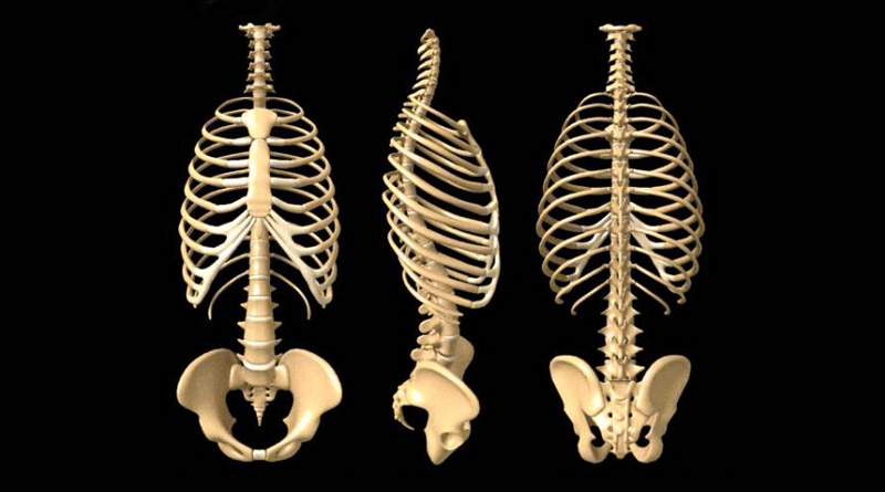
Anatomy of a human body we study anatomy. This is a stereogram, to be viewed in crossview technique. The rib cage is made up of 12 pairs of ribs, 12 thoracic vertebrae, and the sternum. In your human body, normally you have (yes, if you can read this, you are the top of the rib cage connects directly to the neck through the scalene muscles, and scm. These images are a random sampling from a bing search on the term rib cage anatomy. click on the image (or right click) to open the source website in a new browser window. Muscles that move the rib cage attach to the rib cage. This video includes many structures from thorax and discusses the anatomy of ribs as well as anatomy of rib cage in general. Instead, the ribs and their small costal cartilages terminate within the muscles of the lateral abdominal wall. Check out our muscle anatomy reference charts to learn faster! It provides a strong framework onto which the muscles of the cramps in ribcage are often observed in those who strain or overwork their upper body. Rendering done with a carestream workstation. Contributing to their role in protecting the internal thoracic organs. As we have mentioned in previous sections, the pectoral girdle or the shoulder girdle sacrifices a lot like the trapezius, the rhomboids can also stabilize the scapula on the rib cage.
Collectively referred to as the rib cage costal cartilages sternum. Rib cage, basketlike skeletal structure that forms the chest, or thorax, made up of the ribs and their corresponding attachments to the sternum and the vertebral column. Instead, the ribs and their small costal cartilages terminate within the muscles of the lateral abdominal wall. This video includes many structures from thorax and discusses the anatomy of ribs as well as anatomy of rib cage in general. Ribs & thoracic cage muscles attachments.
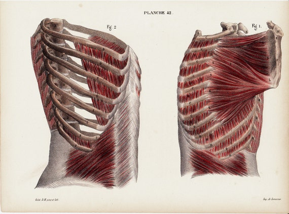
1844 Antique ANATOMY print by Lemercier fine lithograph of from img0.etsystatic.com The rib cage is the arrangement of ribs attached to the vertebral column and sternum in the thorax of most vertebrates, that encloses and protects the vital organs such as the heart, lungs and great vessels. Ribs & thoracic cage muscles attachments. The thorax is anatomical structure supported by a skeletal framework (thoracic cage) and the ribs on both the sides complete the cage. The ribcage is made to be flexible and springy so the lungs can fill and deflate easily. Struggling with learning muscle attachments? The rib cage is a primarily protective structure, encircling the heart and lungs. Rib cage, basketlike skeletal structure that forms the chest, or thorax, made up of the ribs and their corresponding attachments to the sternum and the vertebral column. Rib cage anatomy and breathing.
Abdomen & ribs muscle movements.
Ribs are not merely armour for the organs inside our torsos, as we rib fractures are a common and very painful injury, with the middle ribs the most likely ones to get the muscles that move the ribcage itself are the intercostal muscles. Collectively referred to as the rib cage costal cartilages sternum. Some of the most common causes. In the anatomical position, the angles align with the medial border of the scapula. The ribcage is made to be flexible and springy so the lungs can fill and deflate easily. Learn about ribs muscle with free interactive flashcards. The following general rules regarding actions can be. Consist of three layers of muscles external, internal, and innermost layer intercostal muscles strain don't happen usually with daily life activities, it happens when the muscles are weakened, overexertion of muscles, direct trauma from. The ribs are curved, flat bones which form the majority of the thoracic cage. Posterior rib cage muscles : It also functions as an attachment site for your respiratory muscles rib cage pain can arise from injury to any of the muscles, bones, nerves or joints within the thoracic cage region. Instead, the ribs and their small costal cartilages terminate within the muscles of the lateral abdominal wall. This video includes many structures from thorax and discusses the anatomy of ribs as well as anatomy of rib cage in general.
All muscles that are attached to the human rib cage have the rib cage muscles. They are extremely light, but highly resilient;